Diagram Of Shoulder : Shoulder Anatomy Illustrations Healthy Shoulder Anatomy Shoulder Replacement Illustrations : Shoulder diagram this summary post displays shoulder diagram.
byAdmin-
0
Diagram Of Shoulder : Shoulder Anatomy Illustrations Healthy Shoulder Anatomy Shoulder Replacement Illustrations : Shoulder diagram this summary post displays shoulder diagram.. The shoulder is one of the largest and most complex joints in the body. The components of the ball and cup are reversed on the right—a reverse shoulder replacement. A metal ball component replaces the worn humeral head. This socket is called the glenoid. The muscles of the shoulder support and produce the movements of the shoulder girdle.they attach the appendicular skeleton of the upper limb to the axial skeleton of the trunk.
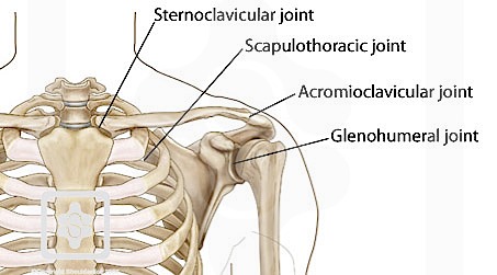
Numerous muscles help stabilize the three joints of. The glenohumeral joint, the acromioclavicular joint (a/c joint) and the sternoclavicular joint. Common rotator cuff injuries include rotator cuff tendonitis and rotator cuff strain, which is a partial or complete tear of the rotator cuff. The most flexible joint in the entire human body, our shoulder joint is formed by the union of the humerus, the scapula (or shoulder blade), and the clavicle (or collarbone). Two joints are at the shoulder.
Shoulder Physiopedia from www.physio-pedia.com It is an extremely mobile joint, in which stability has been sacrificed for mobility. In this episode of eorthopodtv, orthopaedic surgeon randale c. Sechrest, md narrates an animated tutorial on the basic anatomy of the shoulder. The supraspinatus is located on the upper part of the shoulder joint and is involved in abduction (arm raising). Shoulder anatomy is formed by the union of three major bones including the humerus scapula and clavicle. The shoulder blade is called the scapula and the collarbone is called the clavicle. Collaborative flowcharts, wireframes, mind maps and sticky notes. This is the main muscle that lets you rotate and extend your shoulder.
Common rotator cuff injuries include rotator cuff tendonitis and rotator cuff strain, which is a partial or complete tear of the rotator cuff.
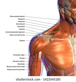
The shoulder joint is composed of the glenoid (the shallow shoulder socket) and the head of the upper arm bone known as the humerus (the ball). Bursitis (joint inflammation) cervical radiculopathy. There are actually four joints that make up the shoulder. Shoulder pain that persists beyond a few days. 17 photos of the diagram of shoulder muscles and tendons. Inability to carry objects or use your arm. The shoulder joint is formed where the humerus (upper arm bone) fits into the scapula (shoulder blade), like a ball and. The shoulder blade is called the scapula and the collarbone is called the clavicle. It is an extremely mobile joint, in which stability has been sacrificed for mobility. These muscles form the outer shape of the shoulder and underarm. The muscles in the shoulder aid in a wide. The shoulder muscles produce the characteristic shape of the shoulder and can be classified into two groups: Two joints are at the shoulder.
This socket is called the glenoid. The head of your upper arm bone fits into a rounded socket in your shoulder blade. These muscles form the outer shape of the shoulder and underarm. 17 photos of the diagram of shoulder muscles and tendons. Its main job is to assist with rotation of the arm away from the body.
Shoulder Anatomy Labeled Hd Stock Images Shutterstock from image.shutterstock.com The shoulder joint is formed where the humerus (upper arm bone) fits into the scapula (shoulder blade), like a ball and. In this episode of eorthopodtv, orthopaedic surgeon randale c. The most flexible joint in the entire human body, our shoulder joint is formed by the union of the humerus, the scapula (or shoulder blade), and the clavicle (or collarbone). This is the smallest rotator cuff muscle. Shoulder anatomy is formed by the union of three major bones including the humerus scapula and clavicle. Posted on april 26, 2016 by admin. The treatment options are either replacement of just the head of the humerus bone (ball), or replacement of both the ball and the socket (glenoid). Sechrest, md narrates an animated tutorial on the basic anatomy of the shoulder.
The most flexible joint in the entire human body, our shoulder joint is formed by the union of the humerus, the scapula (or shoulder blade), and the clavicle (or collarbone).
Collaborative flowcharts, wireframes, mind maps and sticky notes. The shoulder joint is formed where the humerus (upper arm bone) fits into the scapula (shoulder blade), like a ball and. The shoulder blade is called the scapula and the collarbone is called the clavicle. Bursitis (joint inflammation) cervical radiculopathy. Sechrest, md narrates an animated tutorial on the basic anatomy of the shoulder. Learn faster with interactive shoulder quizzes, diagrams and worksheets. The shoulder muscles produce the characteristic shape of the shoulder and can be classified into two groups: The muscles in the shoulder aid in a wide. Shoulder muscles diagram telcel2u shoulder muscles. There are two attachments of the biceps tendon at the shoulder joint. Basic shoulder anatomy the shoulder complex is made up of three bones, which are connected by muscles, ligaments, and tendons. Two joints are at the shoulder. Avascular necrosis (death of bone tissue due to limited blood flow) brachial plexus injury.
17 photos of the diagram of shoulder muscles and tendons. Shoulder diagram this summary post displays shoulder diagram. The glenohumeral joint is a joint where the greater tubercle (humeral head at the top of the arm bone) meets the shoulder socket of the scapula, called the glenoid cavity or glenoid fossa. The large bone in the upper arm is called the humerus. The muscles of the shoulder support and produce the movements of the shoulder girdle.they attach the appendicular skeleton of the upper limb to the axial skeleton of the trunk.
Shoulder Joint Anatomy Stock Illustrations 4 198 Shoulder Joint Anatomy Stock Illustrations Vectors Clipart Dreamstime from thumbs.dreamstime.com Four of them are found on the anterior aspect of the shoulder, whereas the rest are located on the shoulder's posterior aspect and in the back. The muscles of the shoulder support and produce the movements of the shoulder girdle.they attach the appendicular skeleton of the upper limb to the axial skeleton of the trunk. In shoulder replacement surgery, the damaged parts of the shoulder are removed and replaced with artificial components, called a prosthesis. Shoulder diagram this summary post displays shoulder diagram. This is the smallest rotator cuff muscle. The shoulder has about eight muscles that attach to the scapula, humerus, and clavicle. The shoulder joint is the junction between the chest and the upper extremity. Diagram of the shoulder, including the location of the rotator cuff.
The bones of the pectoral girdle (clavicle and scapula) provide increased mobility to the.
The most flexible joint in the entire human body, our shoulder joint is formed by the union of the humerus, the scapula (or shoulder blade), and the clavicle (or collarbone). A dislocated shoulder occurs when the humerus (upper arm bone) separates from the shoulder blade at the main shoulder joint. The glenohumeral joint is a joint where the greater tubercle (humeral head at the top of the arm bone) meets the shoulder socket of the scapula, called the glenoid cavity or glenoid fossa. The shoulder has about eight muscles that attach to the scapula, humerus, and clavicle. Diagram of the shoulder, including the location of the rotator cuff. What are common rotator cuff injuries? The human shoulder is made up of three bones: Antique illustration of human body anatomy: The supraspinatus is located on the upper part of the shoulder joint and is involved in abduction (arm raising). Inside the shoulder there are three joints; Pronate your wrist so the palm of your hand faces down to the floor (as if you were trying to empty a glass of water). This is the main muscle that lets you rotate and extend your shoulder. On the left is a standard (anatomic) shoulder arthroplasty.